OCT: A guide to differentiating lesions
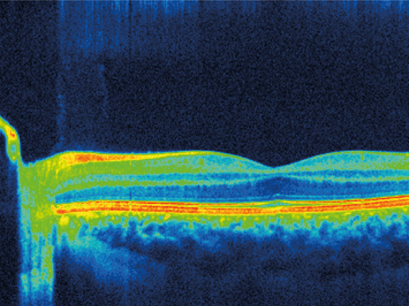
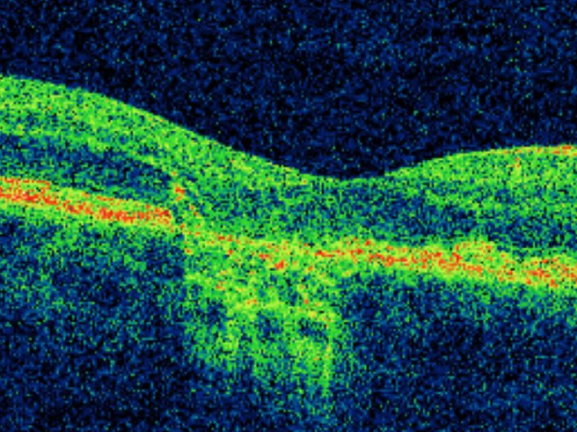
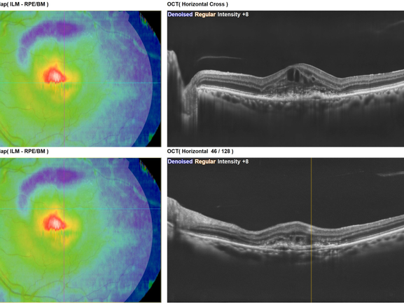
This session will look at some fundamental principles of how to assess each part of an OCT B Scan and differentiate a range of seemingly similar lesions and features to fully understand what pathophysiology is present.
From there, a range of specific eye conditions will be considered and the following we be discussed in detail:
• Fluid and its position
• Leakage, its location and what it means
• AMD – understanding lesion types and differentiating dry, wet and transformational cases
The session will also look at management options, referral criteria and briefly at what further investigative techniques will be used, a brief understanding of those techniques and an introduction to likely treatment options. The introduction will be followed by three case histories, including, AMD, CMO and ERM. Each case will include patient history, patient examination, fundus images and OCT images. These will be used to demonstrate how OCT imaging is relevant in the diagnosis and management of ophthalmic conditions.
CPD Points: 1
Visionstryt credits: 1
Expiry Date: 31/12/2027

Downloads
Accredited by


Approved for


Optometrist (Ocular Examination, Ocular Disease, Standards of Practice)
Dispensing Optician (Standards of Practice, Ocular Examination, Ocular Abnormalities)
Student Optometrist
Student Dispensing Optician


